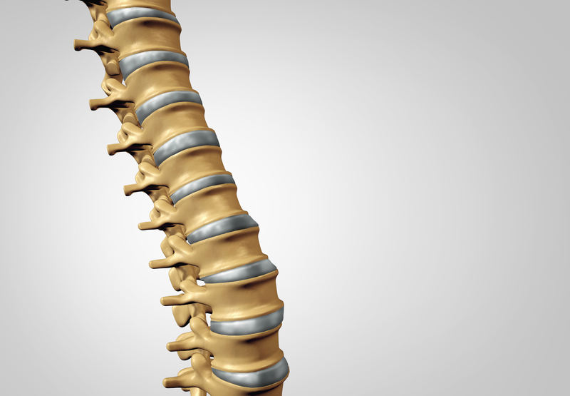
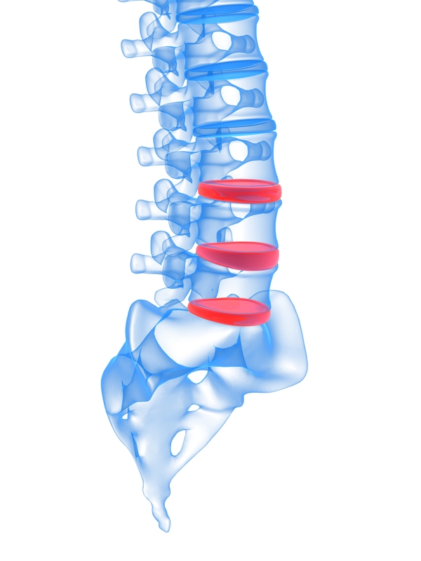
 Your discs play an important role—they help your spine move and also act as a shock absorber. The nucleus, or center, of a disc is made up of a soft jelly-like material. When your discs bulge, they press onto nearby nerves and cause pain. Discography is performed as a diagnostic procedure to help determine the source of pain and how it relates to your discs.
The following might be reasons for a discography:
- Disc degeneration
- Back or neck pain lasting 4 to 6 months
- Back pain that is not responding to other measures (like medication and physical therapy)
How it Works
 Discography is typically performed in the lower two or three lumbar disc levels. During the procedure, a needle is inserted and rests on the outer layer of the disc. A second needle is passed through the first and into the disc’s center. A contrast is then injected into the center of each disc that is being treated. The contrast is viewable through x-ray imaging. In an abnormal disc, the contrast will be seen spreading through the disc’s tears. If the disc is normal, the contrast will stay in the center.
What to Expect
Your doctor will tell you how to prepare for your procedure. There may be eating restrictions and other directions to follow.
X-ray imaging is used to accurately guide the needle to the correct disc. A local anesthetic is used to numb the area. If you experience any pain during the procedure, you may need to describe and rate it in order to help your doctor with your diagnosis.
The procedure itself takes 30 to 45 minutes. You might experience pain at the injection site for a few hours. Other side effects, like infection, are rare. You should be able to return to normal activities in the next day or two.
Your doctor will use the information gained from your discography to determine if surgery or other treatments would be helpful for you.
